Mitochondrial Bioenergetic Remodeling in Type 2 Diabetes: A Comparative Analysis of the Effects of Continuous and Interval Aerobic Training on the Mitochondrial Biogenesis Axis (PGC-1α/TFAM) and Mitochondrial Fusion–Fission Homeostasis (MFN1/DRP1) in the Skeletal Muscle of Diabetic Rats
Keywords:
Type 2 Diabetes, Continuous Aerobic Training, Interval Aerobic Training, PGC-1α, TFAM, MFN1, DRP1, Skeletal MuscleAbstract
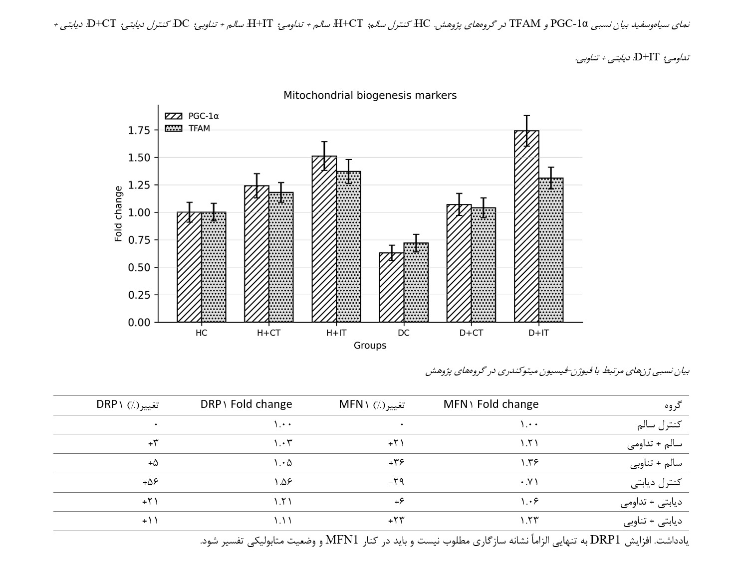
Mitochondrial dysfunction in type 2 diabetes is one of the major mechanisms underlying the reduced metabolic capacity of skeletal muscle. The present study was conducted to compare the effects of continuous and interval aerobic training on the mitochondrial biogenesis axis, including PGC-1α and TFAM, and on the mitochondrial fusion–fission balance, including MFN1 and DRP1, in the gastrocnemius muscle of diabetic rats. In this experimental study, male Wistar rats were assigned, following environmental acclimatization, to six groups: healthy control, healthy + continuous training, healthy + interval training, diabetic control, diabetic + continuous training, and diabetic + interval training (n = 8 per group). Diabetes was induced through a high-fat diet and intraperitoneal injection of streptozotocin. The exercise protocols were performed for eight weeks, five sessions per week. At the end of the intervention, the gastrocnemius muscle was harvested, and the relative expression levels of PGC-1α, TFAM, MFN1, and DRP1 genes were assessed using quantitative real-time polymerase chain reaction (qRT-PCR) and analyzed using the 2^−ΔΔCt method. Type 2 diabetes was associated with decreased expression of PGC-1α, TFAM, and MFN1 and increased expression of DRP1. Both exercise interventions improved mitochondrial indices. Interval training produced a more pronounced increase in PGC-1α and TFAM expression, whereas continuous training demonstrated a more distinct effect on modulating the MFN1 and DRP1 expression profile. The findings suggest that the structure of exercise intensity may activate different pathways of mitochondrial adaptation in diabetic skeletal muscle. Nevertheless, due to the animal-based nature of the study and the absence of certain technical qRT-PCR details in the initial version, the practical interpretation of the findings should be approached with caution and following completion of the methodological reporting.
References
Bustin, S. A., Benes, V., Garson, J. A., Hellemans, J., Huggett, J., Kubista, M., & et al. (2009). The MIQE guidelines: Minimum information for publication of quantitative real-time PCR experiments. Clinical Chemistry, 55(4), 611-622. https://doi.org/10.1373/clinchem.2008.112797
Chavanelle, V., Boisseau, N., Otero, Y. F., Combaret, L., Dardevet, D., Montaurier, C., & et al. (2017). Effects of high-intensity interval training and moderate-intensity continuous training on glycaemic control and skeletal muscle mitochondrial function in db/db mice. Scientific Reports, 7, 204. https://doi.org/10.1038/s41598-017-00276-8
Colberg, S. R., Sigal, R. J., Yardley, J. E., Riddell, M. C., Dunstan, D. W., Dempsey, P. C., & et al. (2016). Physical activity/exercise and diabetes: A position statement of the American Diabetes Association. Diabetes Care, 39(11), 2065-2079. https://doi.org/10.2337/dc16-1728
Fealy, C. E., Mulya, A., Axelrod, C. L., & Kirwan, J. P. (2018). Mitochondrial dynamics in skeletal muscle insulin resistance and type 2 diabetes. Translational Research, 202, 69-82. https://doi.org/10.1016/j.trsl.2018.07.011
Gerhart-Hines, Z., Rodgers, J. T., Bare, O., Lerin, C., Kim, S. H., Mostoslavsky, R., Alt, F. W., Wu, Z., & Puigserver, P. (2007). Metabolic control of muscle mitochondrial function and fatty acid oxidation through SIRT1/PGC-1 alpha. The EMBO Journal, 26(7), 1913-1923. https://doi.org/10.1038/sj.emboj.7601633
Giacomello, M., Pyakurel, A., Glytsou, C., & Scorrano, L. (2020). The cell biology of mitochondrial membrane dynamics. Nature Reviews Molecular Cell Biology, 21(4), 204-224. https://doi.org/10.1038/s41580-020-0210-7
International Diabetes Federation. (2025). IDF Diabetes Atlas (11th ed.). International Diabetes Federation.
Liesa, M., & Shirihai, O. S. (2013). Mitochondrial dynamics in the regulation of nutrient utilization and energy expenditure. Cell Metabolism, 17(4), 491-506. https://doi.org/10.1016/j.cmet.2013.03.002
Little, J. P., Gillen, J. B., Percival, M. E., Safdar, A., Tarnopolsky, M. A., Punthakee, Z., Jung, M. E., & Gibala, M. J. (2011). Low-volume high-intensity interval training reduces hyperglycemia and increases muscle mitochondrial capacity in patients with type 2 diabetes. Journal of Applied Physiology, 111(6), 1554-1560. https://doi.org/10.1152/japplphysiol.00921.2011
Livak, K. J., & Schmittgen, T. D. (2001). Analysis of relative gene expression data using real-time quantitative PCR and the 2^-ΔΔCT method. Methods, 25(4), 402-408. https://doi.org/10.1006/meth.2001.1262
National Research, C. (2011). Guide for the care and use of laboratory animals (8th ed.). National Academies Press. https://doi.org/10.17226/12910
Percie du Sert, N., Hurst, V., Ahluwalia, A., Alam, S., Avey, M. T., Baker, M., & et al. (2020). The ARRIVE guidelines 2.0: Updated guidelines for reporting animal research. PLOS Biology, 18(7), e3000410. https://doi.org/10.1371/journal.pbio.3000410
Pinti, M. V., Fink, G. K., Hathaway, Q. A., Durr, A. J., Kunovac, A., & Hollander, J. M. (2019). Mitochondrial dysfunction in type 2 diabetes mellitus: An organ-based analysis. American Journal of Physiology-Endocrinology and Metabolism, 316(2), E268-E285. https://doi.org/10.1152/ajpendo.00314.2018
Wang, N., Liu, Y., Ma, Y., & Wen, D. (2017). High-intensity interval versus moderate-intensity continuous training: Superior metabolic benefits in diet-induced obesity mice. Life Sciences, 191, 122-131. https://doi.org/10.1016/j.lfs.2017.08.023
World Health Organization. (2024). Diabetes (WHO fact sheet, Issue.
Zheng, L., Rao, Z., Guo, Y., Chen, P., & Xiao, W. (2020). High-intensity interval training restores glycolipid metabolism and mitochondrial function in skeletal muscle of mice with type 2 diabetes. Frontiers in Endocrinology, 11, 561. https://doi.org/10.3389/fendo.2020.00561
Downloads
Publication Timeline
- Submitted
- Revised
- Accepted
Issue
Section
License
Copyright (c) 2025 الهام فنایی (نویسنده); الهام افتخاری قینانی; جمشید بنایی بروجنی (نویسنده)

This work is licensed under a Creative Commons Attribution-NonCommercial 4.0 International License.