بازآرایی زیستانرژی میتوکندری در دیابت نوع ۲: تحلیل تطبیقی اثر تمرینات هوازی تداومی و تناوبی بر محور بیوژنز میتوکندری (PGC-1α/TFAM) و همئوستاز فیوژن-فیسیون میتوکندری (MFN1/DRP1) در عضله اسکلتی رتهای دیابتی
کلمات کلیدی:
دیابت نوع ۲, تمرین هوازی تداومی, تمرین هوازی تناوبی, PGC-1α, TFAM, MFN1, DRP1, عضله اسکلتیچکیده
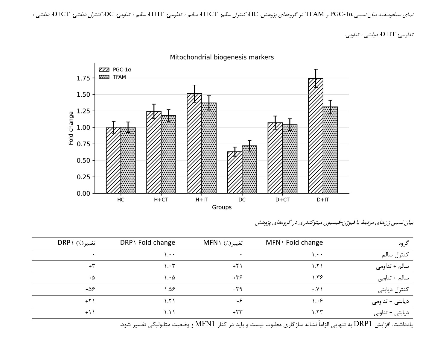
اختلال عملکرد میتوکندری در دیابت نوع ۲، یکی از سازوکارهای مهم کاهش ظرفیت متابولیکی عضله اسکلتی است. پژوهش حاضر با هدف مقایسه اثر تمرینات هوازی تداومی و تناوبی بر محور بیوژنز میتوکندری شامل PGC-1α و TFAM و بر تعادل فیوژن-فیسیون میتوکندری شامل MFN1 و DRP1 در عضله گاستروکنمیوس رتهای دیابتی انجام شد. در این مطالعه تجربی، رتهای نر ویستار پس از سازگاری محیطی به شش گروه کنترل سالم، سالم + تمرین تداومی، سالم + تمرین تناوبی، کنترل دیابتی، دیابتی + تمرین تداومی و دیابتی + تمرین تناوبی تقسیم شدند (n = 8 در هر گروه). دیابت با رژیم غذایی پرچرب و تزریق درونصفاقی استرپتوزوتوسین القا شد. تمرینات به مدت هشت هفته و پنج جلسه در هفته اجرا شدند. پس از پایان مداخله، عضله گاستروکنمیوس استخراج شد و بیان نسبی ژنهای PGC-1α، TFAM، MFN1 و DRP1 با روش qRT-PCR و محاسبه فرمول ۲ به توان منفی ΔΔCt تحلیل گردید. دیابت نوع ۲ با کاهش بیان PGC-1α، TFAM و MFN1 و افزایش DRP1 همراه بود. هر دو مداخله تمرینی شاخصهای میتوکندریایی را بهبود دادند. تمرین تناوبی افزایش برجستهتری در PGC-1α و TFAM ایجاد کرد؛ در حالی که تمرین تداومی اثر مشخصتری بر تعدیل الگوی MFN1 و DRP1 نشان داد. یافتهها نشان میدهد که ساختار شدت تمرین میتواند مسیرهای متفاوتی از سازگاری میتوکندریایی را در عضله اسکلتی دیابتی فعال کند. با وجود این، به دلیل ماهیت حیوانی مطالعه و نبود برخی جزئیات فنی qRT-PCR در نسخه اولیه، تفسیر کاربردی نتایج باید با احتیاط و پس از تکمیل گزارش روششناسی انجام شود.
مراجع
Bustin, S. A., Benes, V., Garson, J. A., Hellemans, J., Huggett, J., Kubista, M., & et al. (2009). The MIQE guidelines: Minimum information for publication of quantitative real-time PCR experiments. Clinical Chemistry, 55(4), 611-622. https://doi.org/10.1373/clinchem.2008.112797
Chavanelle, V., Boisseau, N., Otero, Y. F., Combaret, L., Dardevet, D., Montaurier, C., & et al. (2017). Effects of high-intensity interval training and moderate-intensity continuous training on glycaemic control and skeletal muscle mitochondrial function in db/db mice. Scientific Reports, 7, 204. https://doi.org/10.1038/s41598-017-00276-8
Colberg, S. R., Sigal, R. J., Yardley, J. E., Riddell, M. C., Dunstan, D. W., Dempsey, P. C., & et al. (2016). Physical activity/exercise and diabetes: A position statement of the American Diabetes Association. Diabetes Care, 39(11), 2065-2079. https://doi.org/10.2337/dc16-1728
Fealy, C. E., Mulya, A., Axelrod, C. L., & Kirwan, J. P. (2018). Mitochondrial dynamics in skeletal muscle insulin resistance and type 2 diabetes. Translational Research, 202, 69-82. https://doi.org/10.1016/j.trsl.2018.07.011
Gerhart-Hines, Z., Rodgers, J. T., Bare, O., Lerin, C., Kim, S. H., Mostoslavsky, R., Alt, F. W., Wu, Z., & Puigserver, P. (2007). Metabolic control of muscle mitochondrial function and fatty acid oxidation through SIRT1/PGC-1 alpha. The EMBO Journal, 26(7), 1913-1923. https://doi.org/10.1038/sj.emboj.7601633
Giacomello, M., Pyakurel, A., Glytsou, C., & Scorrano, L. (2020). The cell biology of mitochondrial membrane dynamics. Nature Reviews Molecular Cell Biology, 21(4), 204-224. https://doi.org/10.1038/s41580-020-0210-7
International Diabetes Federation. (2025). IDF Diabetes Atlas (11th ed.). International Diabetes Federation.
Liesa, M., & Shirihai, O. S. (2013). Mitochondrial dynamics in the regulation of nutrient utilization and energy expenditure. Cell Metabolism, 17(4), 491-506. https://doi.org/10.1016/j.cmet.2013.03.002
Little, J. P., Gillen, J. B., Percival, M. E., Safdar, A., Tarnopolsky, M. A., Punthakee, Z., Jung, M. E., & Gibala, M. J. (2011). Low-volume high-intensity interval training reduces hyperglycemia and increases muscle mitochondrial capacity in patients with type 2 diabetes. Journal of Applied Physiology, 111(6), 1554-1560. https://doi.org/10.1152/japplphysiol.00921.2011
Livak, K. J., & Schmittgen, T. D. (2001). Analysis of relative gene expression data using real-time quantitative PCR and the 2^-ΔΔCT method. Methods, 25(4), 402-408. https://doi.org/10.1006/meth.2001.1262
National Research, C. (2011). Guide for the care and use of laboratory animals (8th ed.). National Academies Press. https://doi.org/10.17226/12910
Percie du Sert, N., Hurst, V., Ahluwalia, A., Alam, S., Avey, M. T., Baker, M., & et al. (2020). The ARRIVE guidelines 2.0: Updated guidelines for reporting animal research. PLOS Biology, 18(7), e3000410. https://doi.org/10.1371/journal.pbio.3000410
Pinti, M. V., Fink, G. K., Hathaway, Q. A., Durr, A. J., Kunovac, A., & Hollander, J. M. (2019). Mitochondrial dysfunction in type 2 diabetes mellitus: An organ-based analysis. American Journal of Physiology-Endocrinology and Metabolism, 316(2), E268-E285. https://doi.org/10.1152/ajpendo.00314.2018
Wang, N., Liu, Y., Ma, Y., & Wen, D. (2017). High-intensity interval versus moderate-intensity continuous training: Superior metabolic benefits in diet-induced obesity mice. Life Sciences, 191, 122-131. https://doi.org/10.1016/j.lfs.2017.08.023
World Health Organization. (2024). Diabetes (WHO fact sheet, Issue.
Zheng, L., Rao, Z., Guo, Y., Chen, P., & Xiao, W. (2020). High-intensity interval training restores glycolipid metabolism and mitochondrial function in skeletal muscle of mice with type 2 diabetes. Frontiers in Endocrinology, 11, 561. https://doi.org/10.3389/fendo.2020.00561

دانلود
چاپ شده
ارسال
بازنگری
پذیرش
شماره
نوع مقاله
مجوز
حق نشر 2025 الهام فنایی (نویسنده); الهام افتخاری قینانی; جمشید بنایی بروجنی (نویسنده)

این پروژه تحت مجوز بین المللی Creative Commons Attribution-NonCommercial 4.0 می باشد.